KEY PROJECTS
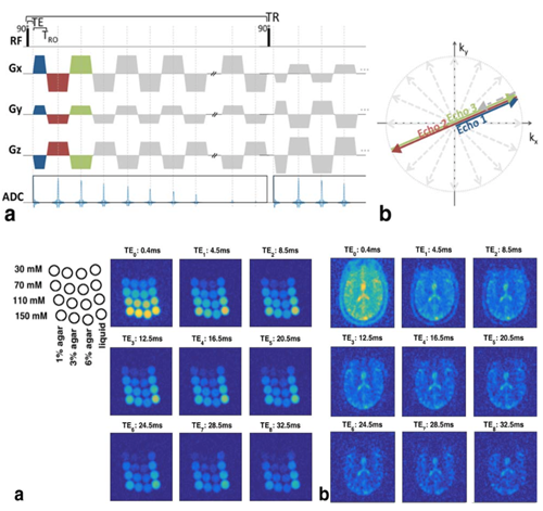
Imaging sodium in the brain
Sodium is critical for neuronal firing and is found in altered distributions in several brain diseases. While Sodium provides the second strongest MR-observable signal in biological tissue, its bulk signal strength is around ~20,000 times less than that of water making it difficult to reliably image, even at high field such as 7 Tesla. Our group has been developing new MRI sequences and post-processing techniques to image sodium and study sodium in different parts of the brain.
For more information please contact Yasmin Blunck.
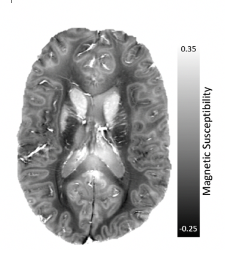
Developing methods for measuring magnetic susceptibility in the brain
MRI can detect changes in the magnetic field produced by small amounts of magnetically interacting substances like iron. In the brain, iron is found in many forms and can interact with brain cells in ways that might lead to neurodegeneration. Researchers at MBCIU are developing new ways of inferring magnetic susceptibility to detect brain iron using a method known as Quantitative Susceptibility Mapping, or QSM. We are also using QSM to study how iron is deposited in the brains of people with neurological diseases like multiple sclerosis and Alzheimer's disease.
For more information please contact Professor Leigh Johnston.
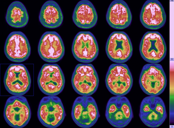
Predicting the onset of dementia using Positron Emission Tomography
Our PET/CT projects include Alzheimer’s research trials that image Amyloid, Tau and glucose to qualify and manage therapeutics that include anti-amyloid, anti tau and others.
We have imaged the largest cohorts in the world for trials A4, EARLY, APAD, 3D, Graduate and many more. We also have been among the first to image across the spectrum with a variety of amyloid and tau PET tracers. As well as other tracers to target MS, Parkinsons disease and schizophrenia. We also use our CT for other purposes such as imaging drill cores fossils, mummies, sheep, dogs, ancient guns, whale ear bones, cane toads, raptors, Tas Devils and for implants and training robots, and for improving the technology by developing PET/CT itself
For more information please contact Rob Williams
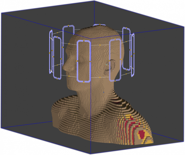
pTx Pulse Design and Simulations
Parallel transmission (pTx) is a promising technology that utilises multiple independent channels in a transmit coil. It offers superior spatial and temporal control over the radiofrequency field and provides extra degrees of freedom to overcome technical hurdles as well as to realise the potential of the ultra-high-field MRI. We at MBCIU are exploring the pTx boundaries and working on pulses for it with uniform excitation and spatial selection. As part of the project, numerical electromagnetic simulations are performed in Sim4Life by ZMT physics solver with realistic computable human body phantoms from the Virtual Population.
For more information please contact Igor Tyshchenko