Melbourne Medical School Researchers Explore the Future of Stent Technology
Researchers from the Melbourne Medical School have published new research exploring a novel device for narrowed or blocked arteries, supporting ongoing efforts to improve patient care and health outcomes.
Stents are tiny mesh tubes used to prop open narrowed or blocked arteries, and they save lives. But leaving a permanent metal implant inside a blood vessel carries long-term risks: reduced flexibility of the vessel wall, slower healing, and clots forming around the device years later. For patients with serious blockages in the leg arteries below the knee, where amputation is a very real threat, better options are needed.
A study recently published in Frontiers in Cardiovascular Medicine, led by Dr Lucas Hatzikostas, Dr Eric Poon, and Professor Peter Barlis from the Department of Medicine, examines one such solution: the resorbable fibrillated scaffold, or RFS.
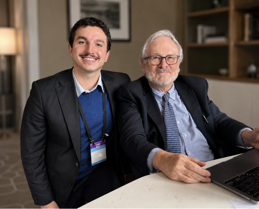
Pictured: Dr Lucas Hatzikostas with his mentor, Professor Patrick Serruys, a pioneer in interventional cardiology and bioresorbable scaffold technology, from the University of Galway.
Think of it as a temporary scaffold — woven from a material the body can break down, it props open the artery long enough for the body's own cells to rebuild the vessel wall, and then dissolves.
Using a high-resolution camera-like probe that travels inside the artery, a technique called optical coherence tomography, combined with computer modelling, Melbourne researchers were the first in the world to image this device from within a living blood vessel. Unlike conventional metal stents, whose small protruding struts disrupt blood flow, the RFS produced a uniform, unobstructed flow environment closer to that of a healthy artery.
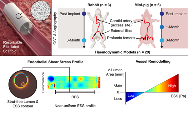
Pictured: The RFS was tested in rabbit and mini-pig arteries over three months. Imaging and computer modelling showed a smooth blood-flow environment linked with vessel wall remodelling. Colour maps show endothelial shear stress (ESS), the frictional force of blood on the artery wall, a key driver of vascular healing. Reproduced from Hatzikostas et al., Frontiers in Cardiovascular Medicine, 2026.
"This device is unique in that it resembles the smooth surface of a healthy artery, restoring more natural conditions inside the vessel," said Dr Lucas Hatzikostas.
The next step is VITAL-IT 1, the first clinical trial testing this novel device in patients with severely reduced blood flow to the legs, where the risk of limb loss is high.