By Dr Jacqueline Healy
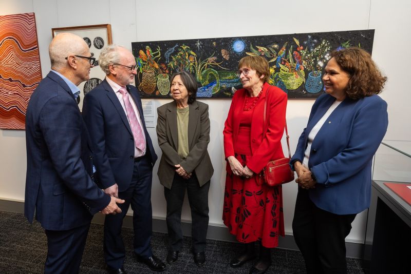
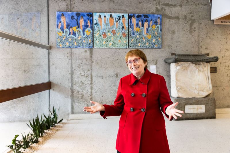
After 58 years in the Brownless Biomedical Library, the Medical History Museum has relocated to a new space on Bouverie Street. The newly refurbished space allows visitors and students to directly engage with the faculty’s extensive collection of objects marking milestones of innovation in medical history.
Here, Director of Museums, Dr Jacqueline Healy reflects on some of the most striking items gifted by distinguished alumni to the museum. Dr Healy reveals a wooden model teaching tool, surreal anatomical drawings, and a silver and bronze microscope.
What stories do they tell, and what do they reveal about the past and future of medicine?
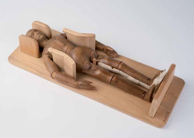
The wooden model that helped break the glass ceiling
At first glance it appears to be a beautifully carved wooden model. However, this object holds a purpose well beyond aesthetics. Resting in a papoose board, the articulated wooden mannequin was designed according to the specifications of Dame Jean Macnamara to illustrate how splinting could prevent deformity in paralysed limbs.
Dame Jean Mcnamara (1899–1968) was a truly pioneering woman. Along with Dame Kate Campbell (1899–1986), she was one of the first female residents at the Royal Children’s Hospital. It took the support of Sir William Upjohn (1888–1979) to get both women appointed after recognising their talent when he worked with them at the Melbourne Hospital.
Dame Macnamara went on to revolutionise the care of children with poliomyelitis, developing an efficient and comprehensive system of care. In collaboration with virologist Sir Macfarlane Burnet, she discovered there was more than one strain of polio virus – an early step towards the development of a vaccine. Dame Campbell specialised in paediatrics, pioneering the field of neonatal medicine.
As an alum of the University of Melbourne, Dame Macnamara chose to give the wooden model she developed to the University to exemplify an aspect of her research for future generations.
Connected by brilliance in surgery and art
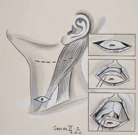
Reminiscent of a Picasso or other surrealist art, this anatomical drawing by Eric Thake (1904–1982) reflects the overlap of medical precision and artistic talent. It came to the museum through surgeon and alum Peter Jones (1922–1995), who commissioned his friend Thake to illustrate surgical techniques.
They are not only beautifully drawn but also demonstrate a style of surgery very close to formal dissection. Professor John Hutson
The remarkable legacy of these illustrations is that they depict a highly invasive technique for treating branchial cleft anomalies requiring extraordinary precision. As Professor John Hutson commented, “They are not only beautifully drawn but also demonstrate a style of surgery very close to formal dissection. This is in stark contrast to current principles of surgery, where we strive to be minimally invasive, avoiding formal dissections unless absolutely necessary.”
Jones graduated from the University of Melbourne with a MBBS in 1945 and went on to become a highly accomplished surgeon. He was the first person to obtain the Australian fellowship in paediatric surgery in 1960 and later edited the world’s first textbook on this new subspecialty as part of the Royal Children’s Hospital’s centenary in 1970.
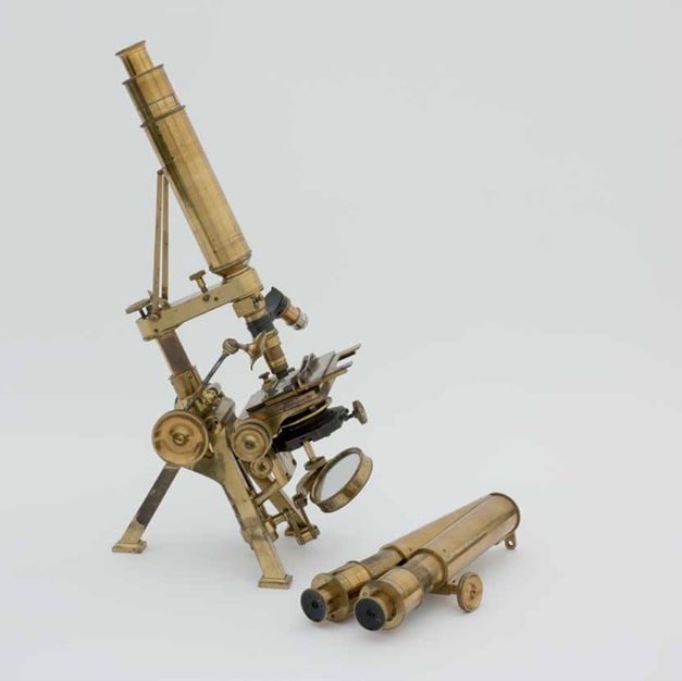
The microscope that built a faculty
This gleaming Powell & Lealand compound monocular and binocular microscope, made of glass and brass, belonged to Professor George Britton Halford (1824–1910), the first Dean of the Faculty of Medicine. The University of Melbourne purchased the microscope in 1864 from the Reverend John Bleasdale (1822–1884), a prominent Catholic clergyman and an active member of the Microscopic Society of Australia. It was donated back to the University by his granddaughter, Mrs Dorothy C Banks, providing a physical link to Professor Halford’s work at the University and the technology of the time.
Professor Halford was appointed the first Chair of Anatomy, Physiology and Pathology at the University of Melbourne in 1862. The medical course commenced with just three students, but by the time the school became a faculty in 1876 there were over sixty. Professor Halford was a progressive advocate for educational access and, as early as 1871, recommended admitting women who passed examinations – a view opposed at the time by Vice-Chancellor A.C. Brownless (1817–1897).
Dr Jacqueline Healy, Director, Museums, Faculty of Medicine, Dentistry and Health Sciences
The Medical History Museum is open
Monday to Friday 10:00am to 5:00pm.
233 Bouverie St, Carlton.
Visit the inaugural exhibition
Cultural Medicine: The Art of Indigenous Healing
from 29/9/2025 to 10/7/2026