New tool discovered to predict best methods of knee cartilage repair in vivo
Dr Carmine Onofrillo and Dr Serena Duchi, from the Department of Surgery, have been the lead authors of a paper based on research resulting in a prediction of what materials will work best to repair knee cartilage in vivo. It is hoped that this new tool will provide new insights to help accelerate future research.
As the ability of human knee cartilage to repair itself is poor, injuries or just regular wear and tear can cause lasting damage to the knee, ultimately leading to painful osteoarthritis.
Along with other labs around the world, the team are developing implantable gel materials loaded with stem cells which might be able to repair the cartilage damage in an early stage, before it develops to full-blown osteoarthritis
Just like any big reno job, building new cartilage requires both construction and destruction. While cells are building new cartilage matrix, the gel is breaking down. The problem is, with all these processes going on at the same time, it’s hard to track what is happening to the original gel, so the research team came up with the idea to use some fluorescent gel to track the breakdown.
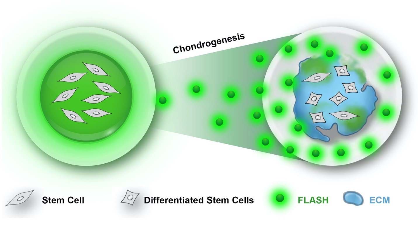
The team decided to add just a small amount of the fluoro gel to our regular GelMA material. As the GelMA breaks down, the fluoro signal leaks into the water. This is what is called the FLASH (Flourescently LAbelled Sensitive Hydrogel) signal.
The resulting paper of this research, FLASH: Fluorescently LAbelled Sensitive Hydrogel to monitor bioscaffolds degradation during neocartilage generation, is where they first show how this FLASH signal closely tracks with the breakdown of the gel (measured by the change in dry weight) in an enzymatic broth, and also when just incubated in saline. When they cultured stem cells in the labelled gels, they measured how the FLASH signal also tracks with the amount of new matrix being made. Confocal images could also clearly distinguish the (green) GelMA from the newly deposited (blue) collagen II matrix:
The team saw the same result for in vitro (petri dish) culture, in a human ex-vivo model (using actual human knee tissue!), and in a rabbit study using three different gel concentrations. This pointed to the fastest degrading gel being the best for cartilage repair.
Overall, they found that just by sampling the culture medium for the FLASH signal in vitro, they could make a solid prediction of what materials will work best to repair knee cartilage in vivo. It is hoped that this new tool will provide new insights to help accelerate future research.
This research was conducted by the following team:
- Dr Carmine Onofrillo, Department of Surgery UoM
- Dr Serena Duchi, Department of Surgery UoM
- Sam Francis, Department of Surgery UoM
- Dr Cathal D. O'Connell, RMIT
- Dr Lilith Caballero-Aguilar, Swinburne University of Technology
- Stephanie Doyle, RMIT
- Dr Zhilian Yue, Australian Research Council (ARC)
- Professor Gordon G. Wallace, Australian Research Council (ARC)
- Professor Peter Choong, Department of Surgery UoM, St Vincent’s Hospital
- Associate Professor Claudia Di Bella, Department of Surgery UoM, St Vincent’s Hospital
Congratulations to Dr Carmine Onofrillo and Dr Serena Duchi and the team!
Article:
Onofrillo C, Duchi S, Francis S, O’Connell CD, Caballero Aguilar LM, Doyle S, et al. FLASH: Fluorescently LAbelled Sensitive Hydrogel to monitor bioscaffolds degradation during neocartilage generation. Biomaterials [Internet]. 2021 Jan 1;264.