The Melbourne Brain Centre Imaging Unit at the University of Melbourne is a Partner of the National Imaging Facility, a National Collaborative Research Infrastructure Strategy (NCRIS) capability.
![]()
![]()
STAFF

Professor Leigh Johnston is the Director of the Melbourne Brain Centre Imaging Unit and a member of the Department of Biomedical Engineering within the School of Chemical and Biomedical Engineering. Her primary research focus is medical imaging, in particular Magnetic Resonance Imaging. She holds an honorary appointment at the Florey Institute of Neuroscience and Mental Health. She coordinates the Melbourne Brain Centre Imaging Unit, with research programs utilising the Siemens 7T and Siemens PET/CT clinical systems on the Parkville campus. Her expertise in MRI spans from acquisition sequences to image analysis and applications. Prior to her appointment at The University of Melbourne, Leigh was a postdoctoral researcher at the Howard Florey Institute (Melbourne), York University (Canada), and the Université catholique de Louvain (Belgium).
Carly Beveridge is our Research Nurse working in the MBCIU to support participants through their imaging appointments to ensure they have best possible care and experience, always working to put study participants at ease.
Carly has come into the MBCIU with several years of nursing experience in Emergency Departments where she has developed and refined her nursing skills. As a nurse she is applying her knowledge and experience to make a great difference to our operations, participants and researchers alike.
Carly has always been fascinated about how the brain and body work and is enjoying learning and working on the wide of research topics and projects supported at the MBCIU.
Dr. Yasmin Blunck is a Doreen Thomas Postdoctoral Fellow in the Department of Biomedical Engineering within the School of Chemical and Biomedical Engineering. She is affiliated with the Melbourne Brain Centre Imaging Unit where she develops new methods on the 7T research MRI. Her current research focus is on Sodium Imaging with a particular interest in image acquisition, i.e. sequence development, and reconstruction techniques.
Prior to her PhD at the University of Melbourne where she graduated in 2018, she received a Bachelor of Science in Biomedical Engineering from the University of Applied Sciences Stralsund, Germany, and a Master of Science in Biomedical Computing from the Technical University of Munich, Germany.
During her academic degrees, she completed internships at the Montreal Neurological Institute, Fraunhofer Mevis, Siemens Healthineers and IBM research where she worked on various projects in biomedical imaging.

Associate Professor Katie Davey ![]()
Dr. Katie (Catherine) Davey is an Associate Professor in the Biomedical Engineering Department at the University of Melbourne. Katie's primary research areas are in functional MRI - the acquisition and analysis of a series of low resolution magnetic resonance images to understand brain function - and spike timing dependent synaptic plasticity (STDP) - the process by which connected neurons adapt connection strengths during learning. Katie's research uses advanced signal processing methods, in conjunction with simulation and modelling techniques, to mathematically and programmatically model cortical processes and gain insight into how we perceive and process sensory information.
Katie has additional collaborations, such as with Imperial College of London to investigate the encoding and storage of location information by place cells using calcium imaging, and with Florey Institute of Neurosciences in modelling the neural pathways for bowel disease.
Katie completed her doctoral research in functional MRI connectivity, which is a field of research that analyses a series of low resolution MRI images to identify how brain regions cooperate to achieve sensory and perception tasks. After completing her Ph.D. Katie worked at the Defence Science Technology Organisation, modelling pilot cognition and aircraft control. She then worked in finance, modelling and predicting the movement of stock prices on the S&P500.

Dr. Georgia Giannakis is the MBCIU Platform Manager and brings experience from a range of medtech, diagnostic and medical research roles. Georgia has an extensive background in business development and research facility operations, including client, finance and contract management. In her most recent role, Georgia was the Clinical Development Manager at Bionic Vision Technologies Pty Ltd, the company that was established from the Bionic Vision Australia consortium where she was involved in the coordination of project activities leading up to the clinical trial of the Australian bionic eye. Her role encompassed project management, risk and document management associated with the clinical trial, (ethics submissions, risk management, device specification documentation, regulatory approvals, associated SOPs and GCP trial documentation).
Within the MBCIU Georgia is available to assist with all service enquiries, contract and business operational matters.

Rebecca Glarin is the Supervisor Radiographer for the 7T MRI Imaging service. She brings with her a wealth of knowledge regarding the safe use of MRI for research and clinical services with over 20 years of experience in the field.
Her position is responsible for assisting in protocol development and liaising with facility users to provide optimal imaging protocols specifically developed to address the group’s research aims. The position acquires scans involving human participants on the 7T MRI system and is responsible for the delivery and testing of MRI sequences for both human and phantom-based research. In addition, it is also responsible for data quality control, data curation and management of the MRI bookings system and DaRIS data distribution system, and responsible for the development of safe operating procedures at the facility.
Rebecca is a PhD candidate in the Baker Department on Cardiometabolic Health at the University of Melbourne. Her current work is the investigation of sympathetic drive by combining microneurography with ultra-high field functional MRI. The primary aim is to identify subcortical nuclei and the autonomic circuitry responsible for blood pressure control and thermoregulation.

Lauren is the MBCIU Chief Nuclear Medicine Technologist bringing over a decade of experience in nuclear medicine with a specialized focus in positron emission tomography (PET). Lauren has extensive clinical and research experience having worked at Monash Health and Monash Biomedical Imaging at Monash University using hybrid imaging modalities including a PET/MR system. Lauren oversees the day-to-day technical operations of the imaging service, supports protocol development for research studies to ensure the highest standards of image quality, radiation safety, and participant care. Lauren’s key areas of interest include the use of PET imaging to develop personalised healthcare approaches that improve disease detection and treatment. She is particularly focused on leveraging technological advancements to improve both the participant experience and data quality, while supporting early-career researchers in their use of PET imaging to inspire the next generation of nuclear medicine innovation.
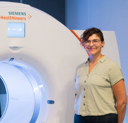
Simon is the Siemens Healthineers on-site scientist dedicated to foster R&D collaborations around 7T MRI with the MBCIU academic team. While supporting the research activity on technical topics, he is driving the research projects on parallel transmission (pTx) pulse design and sequence development to improve image quality and unlock the full potential of 7T MR systems.
As for his academic background, he holds an Engineering degree from Ecole des Mines (France), a Research Master degree in MRI of the spinal cord from Polytechnique Montreal (Canada), a Ph.D. in 7T MRI from Aix-Marseille University (France) and he completed a Postdoc at University Hospital Erlangen (Germany) in pTx pulse design at 7T and in 0.55T MRI of the lung. He was elected as Junior Fellow of the International Society for Magnetic Resonance in Medicine (ISMRM) in 2022, before joining Siemens Healthineers in June 2022.

Sophie manages the Optically Pumped Magnetometer Magnetoencephalography (OPM-MEG) facility. She supports researchers in experimental protocol development, oversees human OPM-MEG acquisitions to ensure data quality and curation. She is also driving the research projects on non-invasive brain imaging to study cerebellar function, especially its predictive processing. She holds a Medicine degree from Yang Ming Chiao Tung University (Taiwan), a Ph.D. in computational neuroscience from Imperial College London (UK) and completed a Postdoc at University College London and University of Birmingham (UK) on cerebellar MEG. A former consultant neurologist (Taiwan), she is now dedicated to advancing functional neuroimaging research.
Associate Professor Brad Moffat ![]()
Associate Professor Brad Moffat is a medical imaging physicist and chemist with 19 years’ research experience in the Biomedical Imaging fields of MRI and Molecular Imaging. Since graduating from his PhD in 2001 he has made an excellent contribution to this field. He is currently a fellow for The University of Melbourne node of the National Imaging Facility. He has specific expertise in quantitative Ultra High Field (7 Tesla) MR imaging of human subjects and Molecular Imaging biomarker research, development and clinical translation (Royal Melbourne Hospital 2007-14). He has published significant journal articles on fMRI, diffusion MRI, functional diffusion mapping, MR perfusion, MRS, voxel based morphometry, PET and nano-theranostics. NIF fellow. Performs operations management for both 7T MRI and PET/CT. His specific research interests are in the development of UHF MRI technology and molecular imaging analytics. More specifically he conducts research on UHF-MRI pulse sequences, MRI contrast agents, endogenous quantitative UHF MRI, MRI/PET/CT reconstruction techniques, image analytics in clinical trials and molecular imaging informatics.

Tudor is an MRI Radiographer at the MBCIU. He has 10 years of clinical experience in MRI and Interventional Radiology and was previously the Supervising Radiographer in a Cardiac Catheterisation Laboratory. Tudor holds a Bachelor of Applied Science (Medical Radiations – Medical Imaging) and is a Magnetic Resonance Safety Officer (MRSO), (MRSC™).

Dr. Warda Syeda is the Imaging Analysis Fellow at MBCIU, with research interests focused on developing processing and modelling techniques for ultra-high field MRI, such as quantitative susceptibility mapping (QSM) and sodium MRI. Prior to commencing this role, Dr Syeda led pre-clinical neuroimaging at the Melbourne Neuropsychiatry Centre, University of Melbourne. Dr Syeda’s interdisciplinary research aims to facilitate the clinical integration of advanced research in neuropsychiatric disorders, such as autism, schizophrenia and schizotypy disorders in children. She has a background in Electrical and Biomedical Engineering and provides expert contributions in methods development for advanced multivariate image processing and inference.

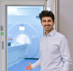
Bahman is a National Imaging Facility (NIF) Fellow in Plant Imaging at the University of Melbourne, contributing to the ARC Centre of Excellence in Plants for Space. His work integrates advanced MRI, PET, and CT technologies to develop next-generation imaging tools for plant systems. With a PhD in Electrical Engineering from the University of Melbourne, Bahman has extensive experience in medical imaging, computational modelling, and AI-driven image analysis. His career includes roles at Bionic Vision Australia (BVA), the Monash Institute of Medical Engineering (MIME), IBM Research Australia, and the Florey Institute of Neuroscience and Mental Health, where he has contributed to diverse areas including MRI physics, neuroimaging, and medical image processing.
PhD CANDIDATES

Joseph Bartlett is undertaking a joint PhD between the University of Melbourne and the University of Birmingham, funded by the Melbourne Research Scholarship. His research focus is on machine learning approaches for improved diffusion MRI. He has completed a Masters of Science in Artificial Intelligence and Machine Learning at the University of Birmingham and was awarded the Computer Science School Prize. He completed his Bachelor of Science in Mathematics at the University of Manchester.

Yining Chen is a PhD student in the Department of Radiology under the Faculty of Medicine, Dentistry and Health Sciences. Her research focuses on the investigation of ultra-high-field MRI biomarkers of chronic traumatic brain injuries. Yining has completed a Bachelor of Diagnostic Radiography at the University of Sydney and a Master of Molecular Imaging Technology at the University of Queensland.

Jamie Elliott is a PhD student in the Department of Biomedical Engineering, University of Melbourne. His research focuses on identifying MRI and PET biomarkers for myalgic encephalomyelitis/chronic fatigue syndrome (ME/CFS). Jamie holds a Bachelor of Pharmaceutical Science from Monash University and conducted his Honours research project at the Florey Institute of Neuroscience and Mental Health.

Haoyu Lu is a PhD student in the Department of Biomedical Engineering, University of Melbourne. His research interests include PET image reconstruction, and combination of deep learning and medical imaging, with special focus on inverse problems and scatter correction. Haoyu graduated with a Bachelor of Science degree in Computer Science and Technology from Beijing Normal University.


Shwet Makadiya is a PhD student in the Department of Biomedical Engineering. His research project involves PET image reconstruction for scatter correction. Shwet has a masters in Medical Imaging and Informatics from IIT Kharagpur and he has pre-doctorate fellowship experience from TCS Research and Innovation where he worked on AI for histopathology image processing.

Mara Quach is a PhD student in the Department of Biomedical Engineering, University of Melbourne. Her research is focused on Chemical Exchange Saturation Transfer (CEST) at ultra-high-field. Mara has a Bachelor of Biomedicine and a Master of Engineering from Melbourne University. In Addition to her current post Graduate studies, Mara works for the Alfred Research Alliance as a research assistant on preclinical imaging projects using diffusion-weighted MRI and QSM for application in motor neuron diseases, traumatic brain injury, and other disorders.
Sina Rasti is a PhD student in the Department of Radiology within the Faculty of Medicine, Dentistry and Health Sciences at the University of Melbourne. His research focuses on the quantitative analysis of brain imaging using ultra‑high‑field MRI. Sina holds a medical degree from Isfahan University of Medical Sciences (Iran) and has experience working as a research physician in medical imaging prior to commencing his PhD.
MBCIU Alumni
The Melbourne Brain Centre Imaging Unit at the University of Melbourne is a Partner of the National Imaging Facility, a National Collaborative Research Infrastructure Strategy (NCRIS) capability.
![]()
![]()
Featured content

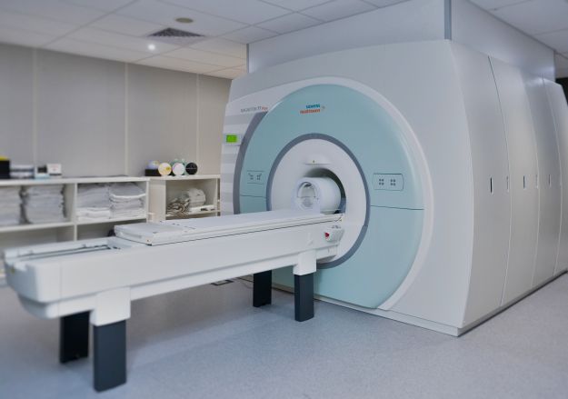
Siemens 7 Tesla MRI Scanner
Our Siemens Magnetom 7T Plus Ultra High Field Magnetic Resonance Imaging scanner is one of only two in Australia. This state of the art scanner provides unprecedented image quality allowing acquisition of non-invasive structural, functional and molecular information at exquisite spatial and temporal resolutions.
The 7T MRI scanner was purchased from Siemens and installed on the ground floor of the Melbourne Brain Centre at Parkville in March 2014. The MBCIU has a Master Research Agreement with Siemens that allows the latest MRI technology to be available on the scanner for research collaborators before they are commercially available.
Research studies on the 7T continue to be primarily neuroimaging focused, however the node has expanded applications with an increase of non-brain studies, given exemplary new functionality with the eye and cervical spine coils, and the flexibility of the knee coil to study wrists and other peripheral limb sections.
The facility maintains world’s best practice for the following applications and expertise:
- Quantitative structural MRI
- Spectroscopy Imaging
- Quantitative Susceptibility Mapping (QSM)
- Sodium MRI
- Neuro-Vascular Imaging (Time of Flight and SWI)
- Functional MRI (fMRI)
- Ultra-high field MRI Safety (Accredited MRI safety officer and expert)
The MBCIU is the University of Melbourne Node of the National Imaging Facility (NIF, https://anif.org.au/). This facility has funded the purchase and operations of the Human 7T MRI and PET/CT scanners via the Australian Government NCRIS program, and Victorian Government matching funds. The purpose was to provide Australian scientists with world-class neuroimaging facilities.
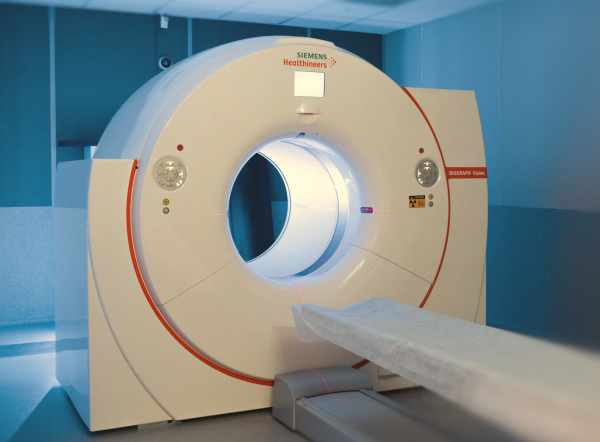
Siemens PET-CT Scanner
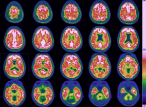
The Imaging Unit within the Melbourne Brain Centre at Parkville houses a state-of-the-art PET-CT scanner, which has been funded under the umbrella of a successful VSA bid submitted by the Victorian Biomedical Imaging Consortium (VBIC). Radiopharmaceuticals can be scanned in order to study tissue function and metabolism and novel biomarkers are being used to study brain disorders such as Alzheimer's disease. The system can provide time-of-flight measurements and has an extended field of view PET with 128-slice CT.
The Human PET/CT has continued to be well utilised in a broad range of research studies and clinical trials. Optimised reconstruction is developing as a focus for the UOM PET/CT team, using dedicated access to the Siemens e7 toolbox, and in collaboration with CSIRO for standardised tracer uptake computation. A primary objective of this work is to derive metrics by which to evaluate scanner harmonisation in multicentre studies.
OPM-MEG
The Optically Pumped Magnetometer Magnetoencephalography (OPM-MEG) system, funded by ARC LIEF, was procured and installed in 2024. It is the first OPM-MEG system in the Southern Hemisphere and forms part of the MBCIU at the University of Melbourne, giving researchers access to an unparalleled suite of imaging technology for structural and functional brain research, all within at single world-class facility.
MEG enables continuous recordings of ongoing brain activity with millisecond-level temporal resolution. OPM-MEG uses small, lightweight sensors that operate at room temperature and can be positioned close to the scalp, enhancing both signal strength and spatial resolution. This wearable and flexible configuration enables the recording of brain activity during naturalistic and interactive conditions, including movement, speech, and sensory tasks, enabling the study of cognition and brain function in more ecologically valid settings. OPM-MEG is non-invasive and well suited for research in neurological, psychiatric, or movement disorders.
The system includes 50 Field Line OPM sensors, which can operate as 50 single-axial sensors or be configured to form 100 dual-axial or up to 150 tri-axial channels. This technology can be used to investigate auditory and visual processing, cerebellar activity, neurobiological markers in psychotic disorders, and the development of next-generation brain–computer interfaces.
The peripheral OPM-MEG equipment include:
- ProPixx visual projector (1440 Hz refresh rate)
- SOUNDPixx auditory stimulation
- RESPONSEPixx /MRI Dual handheld 5-button fiber-optic response box
- Stimulus delivery computer (Psychtoolbox and PsychoPy)

The Melbourne Brain Centre Imaging Unit at the University of Melbourne is a Partner of the National Imaging Facility, a National Collaborative Research Infrastructure Strategy (NCRIS) capability.
![]()
![]()
MELBOURNE BRAIN CENTRE IMAGING UNIT
To contact us, please fill in our Enquiries Form.
We will direct your enquiry to the most appropriate person and respond within 1-2 working days.
Address:
Melbourne Brain Centre Imaging Unit
Ground Floor - Kenneth Myer Building
30 Royal Parade, The University of Melbourne.
Email:
Map
Volunteers:
You can help us by volunteering for clinical and research studies. The MBCIU is involved in numerous trials involving many debilitating diseases. Contact us to learn more about how you can help shape the future of research at the University of Melbourne.